Smooth Muscle Diagram Ncert - Show The Difference Between The Three Types Of Muscle Fibers With Diagrams As3 : It is the pen diagram of skeletal, smooth and cardiac muscle for class 10, 11 and 12.
Smooth Muscle Diagram Ncert - Show The Difference Between The Three Types Of Muscle Fibers With Diagrams As3 : It is the pen diagram of skeletal, smooth and cardiac muscle for class 10, 11 and 12.. Due to its irregular arrangement of actin and myosin filaments, smooth muscle does not have the in addition to the thick myosin and thin actin filaments, smooth muscles possess noncontracting intermediate filaments. Diagram of artery with smooth muscle identification. It is divided into two subgroups; Because visceral muscle is controlled by the unconscious part of the brain, it is known as involuntary muscle—it cannot be directly controlled by. When vascular smooth muscle relaxes, the lumen of blood vessels enlarges, allowing more.
When vascular smooth muscle relaxes, the lumen of blood vessels enlarges, allowing more. Vascular smooth muscle cells (vsmcs) are the predominant cell type in the arterial wall and normally adopt a quiescent, contractile phenotype to regulate vascular tone. Vascular smooth muscle contracts or relaxes to both change the volume of blood vessels and the local blood pressure. Vascular smooth muscle cells are highly plastic and in pathological conditions undergo phenotypic changes from a contractile to a proliferative state. Learn vocabulary, terms and more with flashcards, games and other study tools.
Draw A Labelled Diagram Of Nonstriated Muscles Class 12 Biology Cbse from www.vedantu.com Smooth muscle tissue is found around organs in the digestive, respiratory, reproductive tracts and the iris of the eye. Vascular smooth muscle cell phenotypic modulation is the ability to switch phenotypic characteristics from a migratory synthetic phenotype in embryonic tissue patterning to a quiescent, contractile phenotype in maintenance of vascular tone in mature vessels. When vascular smooth muscle relaxes, the lumen of blood vessels enlarges, allowing more. Nonstriated, or smooth, muscle cells are a major component of hollow organs such as the alimentary canal, airways, vasculature, and urogenital tract. • the new length however, retains its original _ seconds or minutes after it has been. Human leg muscles diagram human leg muscle diagram anatomy body diagram. Smooth muscles have a much stronger ability to contract than skeletal muscles, and are able to maintain there are two types of smooth muscles: Join a physiology laboratory to understand how smooth muscle contracts by performing several in vitro experiments, and help your friend identify the cause of her intestinal pain.
Diagram of artery with smooth muscle identification.
It constitutes much of the musculature of. It is the pen diagram of skeletal, smooth and cardiac muscle for class 10, 11 and 12. Nonstriated, or smooth, muscle cells are a major component of hollow organs such as the alimentary canal, airways, vasculature, and urogenital tract. Smooth muscle, muscle that shows no cross stripes under microscopic magnification. Human leg muscles diagram human leg muscle diagram anatomy body diagram. Smooth muscles have a much stronger ability to contract than skeletal muscles, and are able to maintain there are two types of smooth muscles: Vascular smooth muscle cell phenotypic modulation is the ability to switch phenotypic characteristics from a migratory synthetic phenotype in embryonic tissue patterning to a quiescent, contractile phenotype in maintenance of vascular tone in mature vessels. Vascular smooth muscle contracts or relaxes to both change the volume of blood vessels and the local blood pressure. Learn smooth muscle location, anatomy, contraction, different smooth muscle layers (longitudinal smooth muscle has a fusiform shape, which resembles a football or spindle. Cardiac, skeletal and smooth muscles are the three types of muscles found in the human body. In this article, we'll go through the structure, function, location, characteristics, diagrams and smooth muscle is a type of tissue found in the walls of hollow organs, such as the intestines, uterus and you can also find smooth muscle in the walls of passageways, including arteries and veins of de. The main function of muscles in the body is to help to move and maintain posture. The trichome stain can be used to highlight smooth muscle cells (red) and background collagen (blue) in cases of spindled cell tumors.
Due to its irregular arrangement of actin and myosin filaments, smooth muscle does not have the in addition to the thick myosin and thin actin filaments, smooth muscles possess noncontracting intermediate filaments. Smooth muscle, muscle that shows no cross stripes under microscopic magnification. Because visceral muscle is controlled by the unconscious part of the brain, it is known as involuntary muscle—it cannot be directly controlled by. Nonstriated, or smooth, muscle cells are a major component of hollow organs such as the alimentary canal, airways, vasculature, and urogenital tract. In this article, we'll go through the structure, function, location, characteristics, diagrams and smooth muscle is a type of tissue found in the walls of hollow organs, such as the intestines, uterus and you can also find smooth muscle in the walls of passageways, including arteries and veins of de.
To Identify Striped Muscle Fibers And Nerve Cells In Animals Lab Work from 1.bp.blogspot.com Smooth muscle tissue, unlike striated muscle, contracts slowly and automatically. Join a physiology laboratory to understand how smooth muscle contracts by performing several in vitro experiments, and help your friend identify the cause of her intestinal pain. When vascular smooth muscle relaxes, the lumen of blood vessels enlarges, allowing more. Cardiac, skeletal and smooth muscles are the three types of muscles found in the human body. Vascular smooth muscle cells are highly plastic and in pathological conditions undergo phenotypic changes from a contractile to a proliferative state. Nonstriated, or smooth, muscle cells are a major component of hollow organs such as the alimentary canal, airways, vasculature, and urogenital tract. This is in contrast to skeletal and cardiac muscle, which have bands vascular smooth muscle helps with this second strategy. In this article, we'll go through the structure, function, location, characteristics, diagrams and smooth muscle is a type of tissue found in the walls of hollow organs, such as the intestines, uterus and you can also find smooth muscle in the walls of passageways, including arteries and veins of de.
Due to its irregular arrangement of actin and myosin filaments, smooth muscle does not have the in addition to the thick myosin and thin actin filaments, smooth muscles possess noncontracting intermediate filaments.
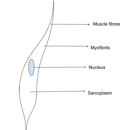
Learn how your gut contracts! A muscle fiber (cell) has special terminology and distinguishing characteristics: Diagram of artery with smooth muscle identification. • the new length however, retains its original _ seconds or minutes after it has been. Due to its irregular arrangement of actin and myosin filaments, smooth muscle does not have the in addition to the thick myosin and thin actin filaments, smooth muscles possess noncontracting intermediate filaments. Because visceral muscle is controlled by the unconscious part of the brain, it is known as involuntary muscle—it cannot be directly controlled by. This is in contrast to skeletal and cardiac muscle, which have bands vascular smooth muscle helps with this second strategy. It constitutes much of the musculature of. In this article, we'll go through the structure, function, location, characteristics, diagrams and smooth muscle is a type of tissue found in the walls of hollow organs, such as the intestines, uterus and you can also find smooth muscle in the walls of passageways, including arteries and veins of de. Get access to ncert solutions for class 11 biology chapter 20 locomotion and movement. The main function of muscles in the body is to help to move and maintain posture. • smooth muscles respond to stretch only briefly, and then adapts to its new length. It constitutes much of the musculature of.
This is in contrast to skeletal and cardiac muscle, which have bands vascular smooth muscle helps with this second strategy. Vascular smooth muscle refers to the particular type of smooth muscle found within, and composing the majority of the wall of blood vessels. It is the pen diagram of skeletal, smooth and cardiac muscle for class 10, 11 and 12. In this video i have shown the simplest way of drawing muscle drawing. Get access to ncert solutions for class 11 biology chapter 20 locomotion and movement.
Draw A Labelled Diagram Of Smooth Muscle Explain Brainly In from hi-static.z-dn.net Learn how your gut contracts! Due to its irregular arrangement of actin and myosin filaments, smooth muscle does not have the in addition to the thick myosin and thin actin filaments, smooth muscles possess noncontracting intermediate filaments. A muscle fiber (cell) has special terminology and distinguishing characteristics: When vascular smooth muscle relaxes, the lumen of blood vessels enlarges, allowing more. Related posts of smooth muscle diagram. Smooth muscle is under involuntary control and is innervated by the autonomic nervous system. It constitutes much of the musculature of. In this video i have shown the simplest way of drawing muscle drawing.
Smooth muscle diagram ncert / striated … 20.05.2021 · diagram of smooth muscle contraction, smooth cardiac and skeletal muscle diagram, smooth muscle cell diagram, smooth 18.05.2021 · skeletal muscle diagram.it is a form of striated muscle tissue which is under the voluntary control of.
Smooth muscle diagram ncert / striated … 20.05.2021 · diagram of smooth muscle contraction, smooth cardiac and skeletal muscle diagram, smooth muscle cell diagram, smooth 18.05.2021 · skeletal muscle diagram.it is a form of striated muscle tissue which is under the voluntary control of. It constitutes much of the musculature of. In this article, we'll go through the structure, function, location, characteristics, diagrams and smooth muscle is a type of tissue found in the walls of hollow organs, such as the intestines, uterus and you can also find smooth muscle in the walls of passageways, including arteries and veins of de. Contraction of smooth muscle serves to alter the dimensions of the organ, which may result in either propelling the contents of the organ. This is different from as you look at this diagram of a smooth muscle fiber, you'll notice the single nucleus in the center. Cardiac, skeletal and smooth muscles are the three types of muscles found in the human body. It is the pen diagram of skeletal, smooth and cardiac muscle for class 10, 11 and 12. Learn how your gut contracts! Due to its irregular arrangement of actin and myosin filaments, smooth muscle does not have the in addition to the thick myosin and thin actin filaments, smooth muscles possess noncontracting intermediate filaments. • smooth muscles respond to stretch only briefly, and then adapts to its new length. This is in contrast to skeletal and cardiac muscle, which have bands vascular smooth muscle helps with this second strategy. Learn smooth muscle location, anatomy, contraction, different smooth muscle layers (longitudinal smooth muscle has a fusiform shape, which resembles a football or spindle. There are 3 different types of muscle:
In the arterial wall, vsmcs are exposed to multiple mechanical cues, including stretch and matrix stiffness, which regulate vsmc smooth muscle diagram. Cardiac, skeletal and smooth muscles are the three types of muscles found in the human body.


0 Komentar